

A Mobile Veterinary Ultrasound Service in Brisbane explains
Vet ultrasound group provides a mobile ultrasound service for cats and dogs in Brisbane and surrounding areas. The following article is a guide for general practice veterinarians on how to achieve high quality ultrasound images on their patients.
1. Improve Contact with the skin
Air is a barrier to the propagation of sound waves so it is very important that good contact with the skin is achieved. For dogs and cats this usually requires that patients are clipped; that the skin is cleaned with alcohol; and a generous amount of coupling gel applied.
You rarely get a good image by parting the hair and using a quick squirt of metho!
2. Use Veterinary Pre-sets if available
Using a pre-set provides a good starting point for the examination and reduces the number of changes you need to make to background settings.
3. Select a frequency appropriate to the organ being examined.
Multi-frequency probes allow the operator to change the frequency to obtain the best quality image.
The higher the frequency, the greater the resolution of the image. The lower the frequency, the greater the penetration.
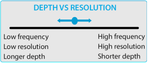
Therefore when imaging a cat; or a superficial organ (eg. a kidney on a large dog) try using a high frequency setting to get the best possible image.
For imaging deeper structures (eg. the liver of an overweight Labrador) you will need a lower frequency probe and as a result may end up with a low resolution image. These patients can be challenging to image!
4. Make use of the Time-Gain Compensation controls
Ever wondered what the slide controls on an ultrasound machine are for?
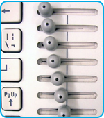
Time gain compensation (TGC) allows the operator to change the image brightness in gradations down the screen.
Ultrasound beams are attenuated as they penetrate tissue: the deeper they travel, the weaker the signal becomes resulting in a darker image.
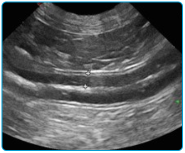
The default position for TGC should be a diagonal from left to right (as seen in the image above).
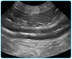
A good way to adjust the TGC is to obtain an image of the liver at the sternum, and alter the TGC controls to obtain an image which is uniform in brightness.5. Make use of Focal Points!
Veterinary ultrasound machines may allow you to change the number and position of focal points on the screen. The ultrasound beams will be concentrated at this level improving image resolution.
Make sure the focal point is placed at the same level as the organ that you are trying to image.

Final Thoughts
Do you have a special interest in diagnostic ultrasound? Improve your skills by performing and observing as many ultrasounds as possible and feel free to contact us for more information. Consider attending a course: the University of Sydney Post-Graduate foundation provides high quality training in veterinary diagnostic ultrasound.
Vet Ultrasound Group practitioners are happy to share their knowledge and experience with general practice veterinarians in Brisbane and surrounds. Happy scanning!

